The Liver Unit of the Department of Surgery at the Milan University, Italy, published in September's issue of HPB journal a study which aimed at determining wether steroid administration may reduce liver injury and improve short term outcome.
The study included 43 patients undergoing liver resection, randomizing the groups to steroid receiving patients and a control group. Patients receiving steroids, were given 500 mg of methylprednisolone preoperatively. Levels of ALT, AST, total bilirubin, AT-III, PT, IL-6, TNF alfa were compared. Length of stay and complications were recorded.
Postoperative serum levels of ALT,AST, total bilirubin, inflammatory cytokines were lower in the steroid group. The incidence of postoperative complications in the control group tended to be higher than the steroid group.
The study concluded that steroid pretreatment represents a potentially important biologic modifier of I/R injury and may contribute to maintenance of coagulant/anticoagulant homeostasis.
Print Post
2007/10/13
Randomized Study of the Benefits of Preoperative Cosrticosteroid Administration on Hepatic I/R Injury

2007/10/10
Surgical Techniques for Liver Transection
Liver resection is still a surgical challenge, due to the high risk of hemorrhage during the transection and the complicated biliary and vascular anatomy of the organ.
The history of the development of surgical techniques for liver resection had it’s main goal, the fight against bleeding. Before 1980’s, liver resection was associated to 10-20% mortality rates.
Currently the in hospital mortality nears 5% in specialized centers. Even though there is a better patient selection, the reduction in blood loss and perioperative transfusions have contributed to the better patient survival. Massive bleeding and transusions not only rise the morbi-mortality risk, but also compromise long term survival after resection of liver tumors, due to the associated immunesuppression, which leads to recurrence.
Finger fracture of the liver parenchyma has been the classic technique for liver resection. During the last 20 years, technological advances have lead to the development of specific instruments such as the compact ultrasonic surgical aspirator, water jet, harmonic scalpel, Ligasure®, and TissuLink®. Another thing that has reduced intraoperative blood loss is the better delineation of the transection plane with the use of IOUS.
Delineation of the Proper Transection Plane
This is not only important to achieve an adequate margin for resection of liver tumors, but also to avoid inadvertent injuries to major hepatic vessels or ducts.
This delineation starts with an adequate knowledge of the computed tomography, to define the different relationships of the tumor and the hepatic veins and biliary pedicles. This can be evaluated transoperatively by IOUS.
Without this knowledge inadvertend injuries may occur during transection, which can lead to massive bleeding or biliary injuries. In some occasions tumor exposition during transection can happen. Generally a 1cm margin is required, but this is still controversial for HCC (Ann Surg 2000; 231: 544-551).
IOUS allows localization of the portal pedicle of each liver segment. Some surgeons perform the liver tattoing with blue dye, injected directly to one portal branch to delineate the segment that is going to be resected.
Techniques of Liver Transection
Finger fracture/clamp crushing
Vacular control is very important to hepatobiliary surgeons. Transection is particularly difficult in cirrhotic livers due to fibrosis. The risk of bleeding is also high in this pathology.
Finger fracture involves crushing the liver parenchyma while under inflow occlusion. This technique was introduced by Lin in 1958 (J Formosa Med Assoc 1958; 57: 742-749). Subsequently was perfected by the introduction of other instruments such as the Kelly clamp (Ann Surg 1974; 180: 285-290). Currently is one of the most widely used techniques today.
Ultrasonic Dissection
In a lot of centers, the use of Cavitron® CUSA (Compact Ultrasonic Surgical Aspirator) has been the standard for liver resection. With this technology, liver parenchyma is fragmented by means of ultrasonic energy and aspirated exposing the vascular and ductal structures, which can be clipped or ligated.
This technique has demonstrated reduction in blood loss, morbidity, when compared with finger crushing. The surgical margin is also wider with this technique (Br J Surg 1996; 83: 117-120).
Water Jet
The water propulsion dissector uses a water jet instead of ultrasonic energy to fragment liver parenchyma and expose vascular and biliary structures. This technique is not as popular as CUSA. Currently only one study showed the results with this technique (Zentralbl Chir 2001; 126: 586-590).
Harmonic Scalpel
More recently, newer technologies have allowed sealing of lesser vessels during liver transection. This technique can be used alone or in conjuction with others, such as finger crushing or CUSA.
It uses ultrasonicaly activated shears to seal small vessels that stand between the vibrating blades. The blades vibrate at 55.5 kHz and can easily dissect parenchyma. The coagulative effect is caused by protein denaturalization that occurs due to destruction of hydrogen bonds and heat generation. The tissue cutting effect derives from a saw mechanism.
It has been used in laparoscopic and open procedures, without reported biliary leaks in Schmidbaier’s study in 2002 (Ann Surg 2002; 235: 27-30).
One disadvantage of this technique is it’s capacity to dissect the liver parenchyma around the hepatic veins, because of its difficulty in controlin large vessels. Even though, it is widely used in laparoscopy.
Ligasure
Another instrumet designed for sealing small vessels using a different principle. Combining compression and bipolar radiofrequency energy, a change in the vessel wall’s collagen and elastin, sealing of vessels as large as 7 mm can be achieved.
Doubts concernig its capacity of sealing large bile ducts persist, and ths was reafirmed in a recent study (World J Surg 2005; 29: 110-112). It is useful for laparoscopic procedures.
TissueLink
A new technology using saline-linked radiofrequency energy has been developed. The saline runs towards the tip of the instrument to link the radiofrequency energy with liver surface, achieving coagulation.
This instrument has a tip that allows transection and vessel sealing simultaneously. Can be used during laparoscopic surgery. You can watch a video here.
Radiofrequency Assisted Liver Transection
Radiofrequency ablation is a good technoque for the treatment of liver tumors. With this technique, a Cool-tip® electrode is inserted in the transection plane serially every 1-2 cm, applying energy for 1-2 minutes, to create coagulated cylinders, that then can be cut by a scalpel.
The advantage of this technique is its simplicity, when compared to the other techniques. One of the potential disadvantages is the sacrifice of liver prenchyma that is coagulated, leaving up to 1 cm of necrotic tissue, which can be critical in cirrhotic patients.
There exists the concern for damage to the hilar structures and hepati veins. The use of this device has to be evaluated. You can watch a video here.
Print Post
2007/10/06
Surgical Management of Colorectal Liver Metastases
The appearance of liver metastases during the evolution of a primary colorectal tumor is a bad prognosis. To prolong the patient's life, many therapies have been proposed, more importantly, chemotherapy.
Today, surgical resection is the only therapeutic option for cure, and attains survival curves at medium and long term, superior to those obtained by other treatments. Resection achieves survival rates of 26 to 45% at 5 years, while it is almost zero in those cases not resected.
Liver resection leads to low morbidity and mortality, close to 1% in specialized centers. It must be emphasized that out of all the patients with colorectal liver metastases, only 10 to 20% can be resected. Today, the challenge is to develop new startegies thatt combine surgical resection with techniques of tumor destruction such as RFA, cryotherapy, portal embolization, and new chemotherapy protocols, with the goal of making those lesions that are initially considered unresectable, resectable.
Principles of Curative Liver Resection
The techniques are divided into 'anatomical' or 'non-anatomical' resections.
Anatomical Resections
They follow the liver segmentation principles described by Couinaud. They are considered as minor, when less than 3 segments are resected, or major, when resection inludes more than 3 segments.
Non-Anatomical Resections
They include the resection of a portion of the liver independently of the liver scisures and glissonian pedicles. They refer mainly to metastasectomies. The liver resection depends on the size of the tumor.
Rules to Respect During Hepatectomy
Independently of the type of liver resection, one must follow these rules in order to avoid postoperative complications:
* Functional liver parenchyma preservation of at least 30% of the total liver mass, to avoid hepatic insufficiency.
* Limit blood loss, in order to avoid transfusions, because it is a known risk factor that favors recurrence.
* Respect the 1 mm margin, to reduce the risk of recurrence.
* Respecto glissonian pedicles destined to the remaining segments, to avoid segmental exclusion, ischemia and necrosis, and biliary fistulas.
Preoperative Patient Evaluation
Before even thinking on performing a liver resection, it is necessary a thorough oncologic examination, seeking other localizations, and also to verify the absence of contra-indications.
Regarding morphological tests, an abdominal US, CT and afterwards a MRI, allow to acquire a better knowledge regarding number of metastases, precise location, relationship with the portal pedicle and the hepatic veins.
Pulmonary localizations must be ruled out systematically, using CT examination. If these lesions are resectable, they dont constitute a contraindication to liver resection. The same goes for all of the extrahepatic metastases.
A colonoscopy is always performed to rule out recurrence, even if the primary tumor has already been resected. If necessary, a bone scan or a brain CT can be performed.
Regarding the blood chemistry, it consists of liver function tests, tumor markers, coagulation profile and clearance of green indocyanine test, which allows to better evaluate liver function, specially in those patients who received neoafjuvant chemotherapy or those with a preexistent liver pathology (hepatitis, cirrhosis).
Valid Indications and New Operative Strategies
A patient with liver metastases limited to one segment without extrahepatic dissemination is always a candidate for liver resection. The challenge for hepatobiliary surgeons is to achieve the necessary resources and strategies that allow the patients' benefit.
The liver surgical progress and the development of new surgical techniques, like RFA, cryotherapy, new radiological interventions such as portal embolization, and new chemotherapy drugs have permitted the elaboration of this new strategies, which make possible curative resections to patients initially considered unresectable. Patients can be divided into four groups:
1. Patients with a voluminous hepatic metastases in which resection leaves an insufficient amount of functional liver parenchyma.
2. Patients with bilobar metastases.
3. Patients with recurrence after resection.
4. Patients with a primary colorectal tumor and synchronous liver metastases.
Patients with a Voluminous Liver Metastases
Two alternative may be offered: Tumor downsizing with systemic or local chemotherapy, and hypertrophy of the future remaining liver (non-tumoral) by portal embolization.
* Downsizing: Bismuth et al, from the Paul Brousse Hospital in France, showed in a study with 53 patients presenting with liver metastases initially non-resectable, that the use of neoadjuvant chemotherapy with 5-fluorouracyl, folinic acid and oxalyplatin, achieved an adequate tumoral downsizing, with similar results as those patients initially resectable. (Ann Surg 1996; 224: 509-522).
* Portal Embolization: When resection is not viable due to insufficient functional liver parenchyma (usually left lobe), surgeons look for hypertrophy of the non-tumoral liver. Once embolization is performed, hypertrophy is evaluated 5-6 weeks after the procedure. If the future remaining liver is >30%, then hepatectomy can be performed.
Patients with Bilobar Metastases
Many therapeutic options have been described:
* Two-stage Hepatectomy including Portal Embolization: The success seen for one stage hepatectomies after portal embolization, lead surgeons to the two stage procedure. This strategy consists on a first stage of metastases resection in the future remnant, and embolization of the tumoral liver. On a second stage, a right hepatectomy is performed, which can be extended to segment 4, after an adequate hypertrophy is achieved.
* Two-stage Hepatectomy: Described by René Adam, consists on the resction of the majority of the metastases in a first stage. Then resection of the remaining lesions after acquiring liver regeneration. In between, patients receive chemotherapy.
* Hepatectomy associated to resection and tumoral destruction by local treatment: Here, the greater lesions are resected and the lesser ones are destroyed locally by either RFA or cryotherapy.
* Two-stage Hepatectomy after neoadjuvant chemotherapy: The goal is to achieve an adequate downsizing of the tumor that allows for a resection in a one-stage or two-stage procedure.
Patients with Recurrence after Resection
Surgery is the only curative option for these patients. It has been shown that patients with a liver recurrence isolated or associated with a resectable extrahepatic metastases, resection of all the tumoral tissue achieves an overall survival similar to patients without recurrence.
Resection of the recurrence turns the clock back to zero. In these cases, tumor destruction by RFA or cryotherapy extends the indications.
Patients with a Primary Colorectal Tumor and Synchronous Metastases
Surgical startegies remain controversial. Those who favor simultaneous resection, and those who oppose it, differ in terms of oncological basis, immunological techniques and patient comfort. Jaeck showed that there was no difference between the two approaches (Chirurgie 1999; 124: 258-263).
Print Post
2007/10/04
Identifying Donors at Risk for Perioperative Complications During LDLT

The University of Rochester and Pennsilvania published a retrospective study this month, regarding the risk factors involving living donor liver transplantation.
It is no secret that this surgery poses some problems. First, the surgery, a hepatectomy is performed on a completely healthy patient, and second, this patient is not benefitted from the procedure. This makes the procedure a technical and ethical challenge.
Recently, during the ESOT Congress, Dr. Rene Adam, from the Paul Brousse Hospital, and memeber of the European Liver Transplant Registry (ELTR), commented on the statistics regarding LDLT. He made special emphasis on the 5 donor deaths to date.
American Journal of Transplantation 2007; 7: 2344–2349
* If you want to read the article you can download it from the "BOX" widget, in the inferior right column.
Print Post Read More......
2007/10/01
ESOT: Adult-To-Adult Living Donor Liver Transplantation Using Dual Grafts

Dr. Chih-Chi Wang from the Department of Surgery at Kaohsiung Hospital, Taiwan, presented at the ESOT Congress, Prague, their experience with dual grafts for living donor liver transplantation.
They presented a prospective study including 150 adult LDLT and 5 dual graft transplantations. According to their results, 2 recipients received 1 right lobe and 1 left lobe, and the other two received two left lobes.
They concluded that dual graft transplantation provides sufficient volume in the recipient without jeopardizing donor safety. They also added that it should be considered and performed by an experienced center when a single donor is rejected based on volumetry requirements.


Print Post
2006/11/03
Photodynamic Therapy for Biliary Tract Cancer
Photodynamic therapy is a method for local destruction of tissue or organisms by generating toxic oxygen and other reactive species using light absorbed by an administered or an endogenously generated photosensitiser.
It is a highly promising treatment for patients with cancer. More recently it has found increasing use as a method of therapy for non-cancerous illnesses. It depends on the exploitation of natural and vital reactions widespread in nature that have driven and preserved life on this planet. Following administration of a photosensitiser or its precursor there is an accumulation or retention in areas of cancer and disease relative to adjacent normal tissue. The photosensitiser is inactive until irradiated by light, following which cellular destruction occurs. The clear attraction of this method is the possibility of some targeting of the disease by drug and by the area irradiated.
Eukaryotic complex multi-cellular organisms have a high degree of metabolic specialisation with a requirement for oxygen and carbon species for oxidative phosphorylation. Yet the generation of reactive oxygen species is an initiator of apoptosis and cell death. Eukaryotic cells have therefore developed methods to resist oxidation. Evolutionary biologists believe that cells overcame this problem by the endosymbosis of mitochondria, a chloroplast like energy complex derived from cellular incorporation of primitive protobacteria. The mitochondria also contain natural photosensitisers called porphyrins, which are necessary for manufacture of the oxygen carrier haemoglobin and the energy transfer system involving the cytochromes.
 These are naturally generated or endogenous photosensitisers able to absorb light and generate toxic oxygen species under certain circumstances. Human cells have protective mechanisms against this toxic oxygen damage and cell death will only occur if a critical threshold of toxic oxygen species is reached and the protective mechanisms associated with the mitochondria are overwhelmed. This threshold effect is important since it can be exploited to allow preservation of normal tissues. Some unfortunate individuals are afflicted with a disease in which excessive photosensitisers are generated by in-born errors of metabolism.
These disorders are called porphyrias and lead to excessive accumulation of porphyrin photosensitisers, which when activated by light in the skin result in profound tissue damage overcoming the cells natural defences. These patients are exquisitely sensitive to light; this is most evident in patients with acute intermittent porphyria who are deficient of porphobilinogen deaminase.
This inherited disorder was highly prevalent in central Europe. The afflicted individuals were exquisitely sensitive to light on the skin and developed excess body hair. In addition, the patients could have red teeth, be unable to venture out during daylight, and have mental
disorders. The discovery that an administered substance could render an organism sensitive to light (photosensitivity) is attributed to Oscar Raab working in Munich. In the winter of 1887–98, Professor Herman von Tappeiner set his student Raab to study the toxicity of aniline dyes on paramecia. Raab recognised that the time to kill was related to the intensity of light in the laboratory.
The most dramatic investigation of photosensitization was by Meyer-Betz who injected himself with a porphyrin compound (haematoporphyrin) and observed the effects of sunlight on his skin.
He published a series of photographs of himself suffering from severe photosensitivity with gross facial oedema and erthyema. He remained sensitive to sunlight for over 2 months. examined many other substances including chlorophyll and called the phenomenon “photodynamic action/photodynamische Erscheinung”.
He demonstrated that a photosensitiser, light and molecular oxygen were necessary. He also suggested that tumours could be treated and some early clinical results were reported in 1905 in combination with the dermatologist Jesionek. They applied eosin to skin tumours and exposed them to white light with some response. Subsequently, the parenteral administration of eosin by the French neurologist, J. Prime, as a treatment for epilepsy resulted in a light induced dermatitis in exposed areas of the skin.
In Berlin during the Second World War, Auler and Banzer demonstrated that photosensitisers tended to localise in tumour and malignant tissue. They injected animals with haematoporphyrin and showed increases of fluorescence in animal cancers.
Dougherty established the modern era working at the Division of Radiation Biology at Roswell Park Memorial Institute, Buffalo, USA. He reported that the systemically injected porphyrin (haematoporphyrin) when activated by red light caused complete eradication of transplanted experimental tumours. He also confirmed the preferential accumulation of the photosensitiser in malignant tissue.
Biology and photophysics of clinical photodynamic Therapy
As stated in the introduction, the destruction of abnormal and diseased tissue after generating or administering a photosensitiser with the direct application of light forms the basis of photodynamic therapy.
The requirements are a photosenitiser, light, oxygen and a substrate to act upon. Each photosensitiser has a specific action spectrum that is the wavelengths of light that are absorbed to produce an excited electronic state.
Photodynamic therapy and tissue destruction requires the excited singlet to undergo spin inversion (intersystem crossing) to the metastable triplet state. The triplet state has a longer lifetime and is generally the reactive state involved in photodynamic therapy. The most usual subsequent action is for the activated triplet photosensitiser to transfer energy to ground state oxygen (which is a triplet) to produce singlet oxygen. This molecule is highly reactive and cannot diffuse far before reacting with other molecules. Major biological targets are membranes that undergo rupture and the cells are destroyed. It has been recently demonstrated that most damage is to the membranes around the mitochondria and the lysosomes. These organelles liberate destructive proteins that induce subsequent cellular destruction.
Selective tumour destruction can be achieved if the photosensitiser is administered in low dosage, since the photosensitiser is photodegraded (in normal tissue) by light irradiation before a critical lethal threshold photodynamic dose is reached. However, tumours that selectively retain a higher concentration of photosensitser are destroyed because this threshold photodynamic dose is achieved and cell death is inevitable.
It is also noticeable that some normal tissues are remarkably resistant to photodynamic therapy. They appear to have a naturally higher photodynamic threshold. This is most apparent in the pancreas. The normal pancreatic acinar cell contains many mitochondria and is
very resistant to oxidative stress, the mitochondria being the ingested ‘chloroplast’. This appears essential since it produces such a cocktail of digestive enzymes and must resist auto digestion. Malignant pancreatic cells have fewer mitochondria and thus fewer toxic oxygen quenching molecules, and are therefore much more sensitive to photodynamic therapy. Selective necrosis of tumours with sparing of normal pancreatic tissue can be demonstrated in experimentally induced tumours.
The most commonly used method of photodynamic therapy is to administer a photosensitiser, intravenously, orally or by local application to an area of abnormality and allow retention and accumulation in the tissue for a period of time prior to irradiation with appropriate wavelength light, usually from a laser. These externally administered photosensitisers tend to accumulate in rapidly growing tissue, blood vessels and the supporting tissue that grows with malignant tumours. Parenteral administration either by injection or by mouth does produce a period of general photosensitivity and accumulation is in stromal supportive tissue rather directly within growing cells.
Cancer
Photodynamic therapy has attracted most interest as a method for the local eradication of cancer. The initial treatment of patients was of large areas of tumour that could not be treated by other means, or had failed conventional therapy following surgery, radiotherapy or chemotherapy. The treatment of advanced cancers is effective in palliation of some of the difficult symptoms associated with blockage of food or air passages.
Recently great interest has been shown in the treatment of biliary and pancreatic cancer. Bile duct or cholangiocarcinoma can be a relatively indolent tumour but treatment with surgery, radiotherapy and chemotherapy is very difficult.
Pilot studies have demonstrated that endoscopic Photofrin-PDT is also effective in the palliative treatment of hilar cholangiocarcinoma. A Phase III trial compared stenting plus PDT (n=20) with stenting alone (n=19) and showed a prolongation of survival by almost a year in stenting plus PDT group. The most recent study of eight patients, who underwent 1-5 treatments, showed that median survival from the date of the first PDT treatment was 276 days, whereas median survival times were 45 and 127 days for bismuth type III and IV tumors treated with stenting alone. The five-year follow-up data of 23 patients showed that median survival after treatment was 11.2 months for MO patients and 9.3 months for all patients. The 1-year, 2-year, 3-year, and 4-year survival rates were estimated to be 47%, 21%, 11%, and 5%, respectively, for MO patients and 39%, 17%, 9%, and 4%, respectively, for all patients. Preliminary results confirm that endoscopic illumination of the biliary tract is safe and effective for inoperable cholangiocarcinoma and can improve cholestasis, performance, and quality of life for an extended period. Since endoscopic PDT appears to be the first approach leading to an improvement in prognosis, it should be offered to patients with inoperable cholangiocarcinoma. Preliminary studies suggest that operative PDT might also improve survival for those patients undergoing surgical resection
Links
Medscape
Journal of Hepatology and Gastroenterology
Journal of Gastroenterology
Pubmed
2006/11/02
Hepatoblastoma
Hepatoblastoma is the most common malignant tumor of the liver in children. Surgery remains the primary means of curative therapy, but the role of chemotherapy in both the adjuvant and neoadjuvant setting has become increasingly important over the past three decades.
EPIDEMIOLOGY
The incidence of hepatoblastoma is highest in infants (11.2 per million) and falls off rapidly, with most cases occurring prior to age 5. SEER reports a male:female ratio of 1.2, however, data from group trials in the U.S. and Europe show a higher male:female ratio, ranging from 1.6 to 3.3 An increased incidence of hepatoblastoma has been reported in Beckwith-Weidemann syndrome (BWS), hemihypertrophy and familial adenomatosis polypi (FAP). However, the extent of risk is difficult to determine due to the rarity of hepatoblastoma.
CLINICAL PRESENTATION
Most patients present with an enlarging abdominal mass. The right lobe is involved three times more commonly than the left, with bilobar involvement seen in 20%-30%, and multicentric involvement in 15%. Serum alpha-fetoprotein (AFP) level is almost always elevated. Bilirubin and liver enzymes are usually normal. Anemia and platelet abnormalities have been reported. Although low platelet counts can occur in hepatoblastoma, thrombocytosis is commonly reported.
The etiology of this finding is unclear, however, the liver is a source of thrombopoietin production and increased thrombopoietin has been reported in hepatoblasoma. There is no clear correlation between AFP and outcome, however, persistence or recurrence of elevated AFP is a sensitive marker of disease. There is a correlation between AFP and extent of disease for all stages, and the rate of decline in AFP with treatment is prognostic. Metastases at diagnoses occur in 10%-20% of patients, with the lung being the predominant site of metastases both at presentation and relapse. Other sites of distant metastases, including brain and bone, are rare and usually occur in the setting of relapsed disease.
IMAGING STUDIES
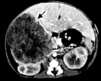 Hepatoblastoma usually appears as a focal or multifocal solid tumor. Stippled or chunky calcifications can be detected in 40%-50% of patients, which is significantly higher than in patients with benign lesions such as hemangiomas and hemangioendotheliomas. Ultrasound in conjunction with color Doppler, a noninvasive modality, is especially useful in young infants. It can assign the tumor to the liver and define its relationship to the portal vein. For the purpose of percutaneous biopsy, either ultrasound or CT guidance can be used to obtain tissue samples for histological analysis.
The imaging work-up used at our institution begins with spiral CT for initial staging of the tumor and for assessing its resectability. It is also used to monitor tumor response to preoperative chemotherapy and search for tumor recurrence. Because pulmonary metastases occur in about 10% of hepatoblastomas, but nonpulmonary metastases are rare, further imaging evaluation recommended at diagnosis should include chest radiography and chest CT to determine if pulmonary metastases are present.
PATHOLOGY/MOLECULAR BIOLOGY
 Hepatoblastoma is classified by histology as epithelial (56%) or mixed epithelial/mesenchymal (44%). Epithelial hepatoblastoma is further broken down to pure fetal (31%), embryonal (19%), macrotrabecular (3%) and smallcell
undifferentiated (3%). The most common mesenchymal elements are osteoid and cartilage. In one study, osteoid made up a small component of 36% of untreated hepatoblastoma, but was increased in treated hepatoblastoma to 82% and composed up to 90% of the tumor area. Multiple cytogenetic abnormalities have been noted in hepatoblastoma with gain of chromosome 20 being the most common, followed by gain of chromosome 2 or 8. Hepatoblastoma is also associated with FAP, and trisomy 20 is a common finding in colon adenomas. Schneider et al. reported a recurring chromosomal aberration, der(4)t(1q;4q), in four hepatoblastoma patients, and two subsequent cases have been reported. Four of the six cases have the same abnormality, der(4)t(1;4)(q12;q34). Structural abnormalities resulting in a gain of material on 1q are also common. The frequency of these abnormalities suggests that they contribute to the etiology of hepatoblastoma.
STAGING
The lack of a uniformly accepted staging system for malignant hepatic tumors in childhood has been an ongoing problem for international comparison. the Intergroup Hepatoma Studies is based on surgical exploration with the completeness of resection and spread of tumor key to staging. Therefore, staging laparotomy and biopsy are essential. The resectability of the primary tumor has no bearing on the staging of hepatoblastomas when distant metastases are present. This staging system is also currently used by the German Cooperative Study Group. The Japanese Society for Pediatric Surgery attempted a classification based on the TNM (tumor, node, metastases), with clinical stage determined by imaging studies prior to surgery. Tumor size, number of involved lobes, regional lymph node involvement, and distant metastases determine stage. The number of liver segments involved and distant metastases were of prognostic significance.
TREATMENT
Surgery
Historically, only complete surgical excision of the primary tumor was felt to correlate with cure. Complete resection of the tumor remains the best hope for long-term survival; however, the advent of effective chemotherapy may permit cure in the presence of initially unresectable or metastatic disease. If at initial laparotomy the tumor appears resectable, reasonably safe attempts should be made to remove the tumor. If the tumor is deemed unresectable and the patient is at high risk for complications, a biopsy is taken and preresection chemotherapy is indicated. Whether a tumor is respectable may be somewhat subjective, since what one surgeon may consider unresectable may be resectable to another. The classic reasons for a tumor to be deemed unresectable include an extremely large tumor that may lead to excessive bleeding, involvement of both the right and left lobes, involvement of major hepatic veins or the inferior vena cava (IVC) and diffuse multifocal disease. After initial chemotherapy, tumor shrinkage allows for easier resection with less blood loss and morbidity. Prior to the use of preoperative chemotherapy, about half of newly diagnosed hepatoblastomas were considered respectable. In more recent reports only about 30% have been considered resectable at diagnosis, since tumors that are likely to result in significant surgical morbidity with resection are now considered unresectable. Chemotherapy has been proven to be effective both in an adjuvant as well as neoadjuvant setting. The use of neoadjuvant chemotherapy has resulted in most nonmetastatic hepatoblastomas being respectable. For those tumors that remain unresectable after chemotherapy or local recurrence, liver transplantation can be an option. In a recent review of the literature on transplantation in hepatoblastoma, Dower and Smith reported on 33 stage III and 39 stage IV tumors. In patients transplanted with stage III tumors, disease-free survival (DFS) with minimum follow-up of one year is 72%. For stage IV tumors the DFS was 54% at 4 to 90 months. For both stage III and IV tumors, the two-year DFS was a minimum of 40%. A recent report by Reyes et al. on 12 patients undergoing transplantation for hepatoblastoma demonstrated a one-, three-, and fiveyear post-transplant survival of 92%, 92%, and 83%. Intravenous invasion, positive hilar nodes, and contiguous spread did not have a significant adverse effect on the outcome. Distant metastasis was responsible for the two deaths. Patients with metastatic disease at diagnosis that resolves with chemotherapy or can be removed surgically can be considered transplant candidates. Resection of pulmonary metastases was most effective when:
A) the primary was resected;
B) metastases develop more than six months post-resection;
C) metastases had a marked response to chemotherapy and AFP dropped to less than 25 ng/ml;
D) resection of metastases occurred soon after the AFP no longer responded to chemotherapy;
E) all gross disease was resected ,
F) there were fewer metastases.
Chemotherapy
The utility of chemotherapy in the treatment of hepatoblastoma began to emerge in the 1970s. Although surgery remains the predominant mode of therapy, chemotherapy has increased the number of resectable hepatoblastomas and decreased the morbidity of surgery.
Other Treatments
Transcatheter arterial chemoembolization or hepatic arterial chemoembolization involves giving chemotherapy and vascular occlusive agents via catheter into the artery supplying the tumor. This offers the advantage of higher tumor concentrations of chemotherapeutic drugs with lower systemic exposure. Occlusion of the artery supplying the tumor resulting in tumor ischemia can be done since tumors receive most of their blood supply from the hepatic artery, while the blood supply to normal liver parenchyma comes primarily from the portal vein. This approach has been used in adults with primary or metastatic liver tumors.
Cryoablation, and more recently radiofrequency ablation, have also been used in the treatment of liver tumors in adults with little experience in children.
J Clin Oncol 1991;9:2167-2176; J Clin Oncol 1993;11:96-99;Eur J Cancer 1998;34:2064-2067;J Pediatr 1998;133:585-586;J Pediatr 1998;132:750;J Pediatr Hematol Oncol 1996;18:446;Am J Roentgenol 1992;158:143-149.
Read More......

