Hepatoblastoma is the most common malignant tumor of the liver in children. Surgery remains the primary means of curative therapy, but the role of chemotherapy in both the adjuvant and neoadjuvant setting has become increasingly important over the past three decades.
EPIDEMIOLOGY
The incidence of hepatoblastoma is highest in infants (11.2 per million) and falls off rapidly, with most cases occurring prior to age 5. SEER reports a male:female ratio of 1.2, however, data from group trials in the U.S. and Europe show a higher male:female ratio, ranging from 1.6 to 3.3 An increased incidence of hepatoblastoma has been reported in Beckwith-Weidemann syndrome (BWS), hemihypertrophy and familial adenomatosis polypi (FAP). However, the extent of risk is difficult to determine due to the rarity of hepatoblastoma.
CLINICAL PRESENTATION
Most patients present with an enlarging abdominal mass. The right lobe is involved three times more commonly than the left, with bilobar involvement seen in 20%-30%, and multicentric involvement in 15%. Serum alpha-fetoprotein (AFP) level is almost always elevated. Bilirubin and liver enzymes are usually normal. Anemia and platelet abnormalities have been reported. Although low platelet counts can occur in hepatoblastoma, thrombocytosis is commonly reported.
The etiology of this finding is unclear, however, the liver is a source of thrombopoietin production and increased thrombopoietin has been reported in hepatoblasoma. There is no clear correlation between AFP and outcome, however, persistence or recurrence of elevated AFP is a sensitive marker of disease. There is a correlation between AFP and extent of disease for all stages, and the rate of decline in AFP with treatment is prognostic. Metastases at diagnoses occur in 10%-20% of patients, with the lung being the predominant site of metastases both at presentation and relapse. Other sites of distant metastases, including brain and bone, are rare and usually occur in the setting of relapsed disease.
IMAGING STUDIES
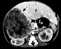 Hepatoblastoma usually appears as a focal or multifocal solid tumor. Stippled or chunky calcifications can be detected in 40%-50% of patients, which is significantly higher than in patients with benign lesions such as hemangiomas and hemangioendotheliomas. Ultrasound in conjunction with color Doppler, a noninvasive modality, is especially useful in young infants. It can assign the tumor to the liver and define its relationship to the portal vein. For the purpose of percutaneous biopsy, either ultrasound or CT guidance can be used to obtain tissue samples for histological analysis.
The imaging work-up used at our institution begins with spiral CT for initial staging of the tumor and for assessing its resectability. It is also used to monitor tumor response to preoperative chemotherapy and search for tumor recurrence. Because pulmonary metastases occur in about 10% of hepatoblastomas, but nonpulmonary metastases are rare, further imaging evaluation recommended at diagnosis should include chest radiography and chest CT to determine if pulmonary metastases are present.
PATHOLOGY/MOLECULAR BIOLOGY
 Hepatoblastoma is classified by histology as epithelial (56%) or mixed epithelial/mesenchymal (44%). Epithelial hepatoblastoma is further broken down to pure fetal (31%), embryonal (19%), macrotrabecular (3%) and smallcell
undifferentiated (3%). The most common mesenchymal elements are osteoid and cartilage. In one study, osteoid made up a small component of 36% of untreated hepatoblastoma, but was increased in treated hepatoblastoma to 82% and composed up to 90% of the tumor area. Multiple cytogenetic abnormalities have been noted in hepatoblastoma with gain of chromosome 20 being the most common, followed by gain of chromosome 2 or 8. Hepatoblastoma is also associated with FAP, and trisomy 20 is a common finding in colon adenomas. Schneider et al. reported a recurring chromosomal aberration, der(4)t(1q;4q), in four hepatoblastoma patients, and two subsequent cases have been reported. Four of the six cases have the same abnormality, der(4)t(1;4)(q12;q34). Structural abnormalities resulting in a gain of material on 1q are also common. The frequency of these abnormalities suggests that they contribute to the etiology of hepatoblastoma.
STAGING
The lack of a uniformly accepted staging system for malignant hepatic tumors in childhood has been an ongoing problem for international comparison. the Intergroup Hepatoma Studies is based on surgical exploration with the completeness of resection and spread of tumor key to staging. Therefore, staging laparotomy and biopsy are essential. The resectability of the primary tumor has no bearing on the staging of hepatoblastomas when distant metastases are present. This staging system is also currently used by the German Cooperative Study Group. The Japanese Society for Pediatric Surgery attempted a classification based on the TNM (tumor, node, metastases), with clinical stage determined by imaging studies prior to surgery. Tumor size, number of involved lobes, regional lymph node involvement, and distant metastases determine stage. The number of liver segments involved and distant metastases were of prognostic significance.
TREATMENT
Surgery
Historically, only complete surgical excision of the primary tumor was felt to correlate with cure. Complete resection of the tumor remains the best hope for long-term survival; however, the advent of effective chemotherapy may permit cure in the presence of initially unresectable or metastatic disease. If at initial laparotomy the tumor appears resectable, reasonably safe attempts should be made to remove the tumor. If the tumor is deemed unresectable and the patient is at high risk for complications, a biopsy is taken and preresection chemotherapy is indicated. Whether a tumor is respectable may be somewhat subjective, since what one surgeon may consider unresectable may be resectable to another. The classic reasons for a tumor to be deemed unresectable include an extremely large tumor that may lead to excessive bleeding, involvement of both the right and left lobes, involvement of major hepatic veins or the inferior vena cava (IVC) and diffuse multifocal disease. After initial chemotherapy, tumor shrinkage allows for easier resection with less blood loss and morbidity. Prior to the use of preoperative chemotherapy, about half of newly diagnosed hepatoblastomas were considered respectable. In more recent reports only about 30% have been considered resectable at diagnosis, since tumors that are likely to result in significant surgical morbidity with resection are now considered unresectable. Chemotherapy has been proven to be effective both in an adjuvant as well as neoadjuvant setting. The use of neoadjuvant chemotherapy has resulted in most nonmetastatic hepatoblastomas being respectable. For those tumors that remain unresectable after chemotherapy or local recurrence, liver transplantation can be an option. In a recent review of the literature on transplantation in hepatoblastoma, Dower and Smith reported on 33 stage III and 39 stage IV tumors. In patients transplanted with stage III tumors, disease-free survival (DFS) with minimum follow-up of one year is 72%. For stage IV tumors the DFS was 54% at 4 to 90 months. For both stage III and IV tumors, the two-year DFS was a minimum of 40%. A recent report by Reyes et al. on 12 patients undergoing transplantation for hepatoblastoma demonstrated a one-, three-, and fiveyear post-transplant survival of 92%, 92%, and 83%. Intravenous invasion, positive hilar nodes, and contiguous spread did not have a significant adverse effect on the outcome. Distant metastasis was responsible for the two deaths. Patients with metastatic disease at diagnosis that resolves with chemotherapy or can be removed surgically can be considered transplant candidates. Resection of pulmonary metastases was most effective when:
A) the primary was resected;
B) metastases develop more than six months post-resection;
C) metastases had a marked response to chemotherapy and AFP dropped to less than 25 ng/ml;
D) resection of metastases occurred soon after the AFP no longer responded to chemotherapy;
E) all gross disease was resected ,
F) there were fewer metastases.
Chemotherapy
The utility of chemotherapy in the treatment of hepatoblastoma began to emerge in the 1970s. Although surgery remains the predominant mode of therapy, chemotherapy has increased the number of resectable hepatoblastomas and decreased the morbidity of surgery.
Other Treatments
Transcatheter arterial chemoembolization or hepatic arterial chemoembolization involves giving chemotherapy and vascular occlusive agents via catheter into the artery supplying the tumor. This offers the advantage of higher tumor concentrations of chemotherapeutic drugs with lower systemic exposure. Occlusion of the artery supplying the tumor resulting in tumor ischemia can be done since tumors receive most of their blood supply from the hepatic artery, while the blood supply to normal liver parenchyma comes primarily from the portal vein. This approach has been used in adults with primary or metastatic liver tumors.
Cryoablation, and more recently radiofrequency ablation, have also been used in the treatment of liver tumors in adults with little experience in children.
J Clin Oncol 1991;9:2167-2176; J Clin Oncol 1993;11:96-99;Eur J Cancer 1998;34:2064-2067;J Pediatr 1998;133:585-586;J Pediatr 1998;132:750;J Pediatr Hematol Oncol 1996;18:446;Am J Roentgenol 1992;158:143-149.


0 Comments:
Post a Comment