Liver biopsy is a very useful tool for diagnosis of chronic liver diseases. It was first described by Paul Elrich in 1883 and later by Menghini in 1958.
Several approaches have been described, each one with its advantages and disadvantages. First, the percutaneous liver biopsy, second the transjugular liver biopsy and third, the laparoscopic liver biopsy.
The transjugular approach was first described by Hanafer and Weiner in 1967. Since then it has been widely accepted as a method of diagnosis with its indications increasing and the quality of the tissue samplings getting better with modifications in the needles.
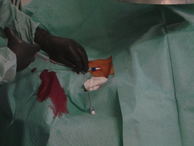 Consists of obtaining liver tissue through a needle that is introduced in the jugular
vein, and through it, a long catheter through the superior vena cava, the right atrium, the inferior vena cava and on through one of the hepatic veins, under fluoroscopic control.
With the catheter in this position, a biopsy needle, 55 cm long, is introduced down to the hepatic vein, and by transfixion of the venous wall, the needle is introduced into the liver parenchyma, to obtain the liver tissue by aspiration or cut.
Measures of pressure gradients in the hepatic veins can be performed to aid in the diagnosis of portal hypertension.
Indications
They are mostly conformed by all the contraindications of percutaneous liver biopsy, and include:
1. Abnormalities of coagulation
a) Platelets: <> 4 seconds over control
2. Massive ascites
3. Vascular tumor or liver peliosis
4. Need to perform other vascular procedure (hemodynamic study and portography)
5. Inability to perform percutaneous liver biopsy
6. Evaluation prior to cardiac and kidney transplant
7. Severe obesity
8. Budd-Chiari Syndrome
Contraindications
1. Hydatid Cyst
2. Biliary dilations (risk of hemobilia)
3. Cardiac anomalies (arrythmias)
Procedure Failure
Arise in a small number of cases (4-8%) and may be operator dependent or inherent to patients anatomy.
1. Impossibility to puncture or cannulate the jugular vein
2. Thrombosis of the right jugular vein
3. Small liver
4. Impossibility to pass the needle through the catheter due to hepatic vein angulation in atrophic livers
5. Early termination of the procedure due to complications
6. Operator’s lack of experience
Quality of the biopsy sample
 The amount of tissue sample is an important factor for an adequate pathological examination. A tissue sample is considered adequate when at least 6 to 8 portal triads are obtained.
The operators experience is important to tissue sampling. Multiple reports describe success rates of 97.1% when the operator had performed more than 100 procedures.
Multiples studies have compared percutaneous and transjugular tissue samples and have fond no significant difference between the two methods.
Complications
Minor complications can be seen in 1-15% of the cases. They include bleeding from the vein puncture site , hematoma, localized pain, and less frequently, Horner Syndrome,
paresthesia, dysphonia and cardiac atrial arrhythmia caused by catheter manipulation on the right atrium, which ceases spontaneously in most cases.
Major complications arise in 1 to 3% of the cases perforation of the hepatic capsule, cholangitis, and intra-peritoneal bleeding. The procedure related mortality is about 0.2 to 0.3%.
Gastroenterology 1990; 99: 1396; Hepatology 1997; 25: 118; Gastrointest Endosc 1999; 50: 536-542; N Eng J Med 2001; 344: 495-500; Semin Liver Dis 1995; 15: 340-359.


0 Comments:
Post a Comment